


 location:Home-Support-Living Cell Imaging Analysis System-EOS Unlabeled cell segmentation ONE BY ONE

A neat and smooth workflow
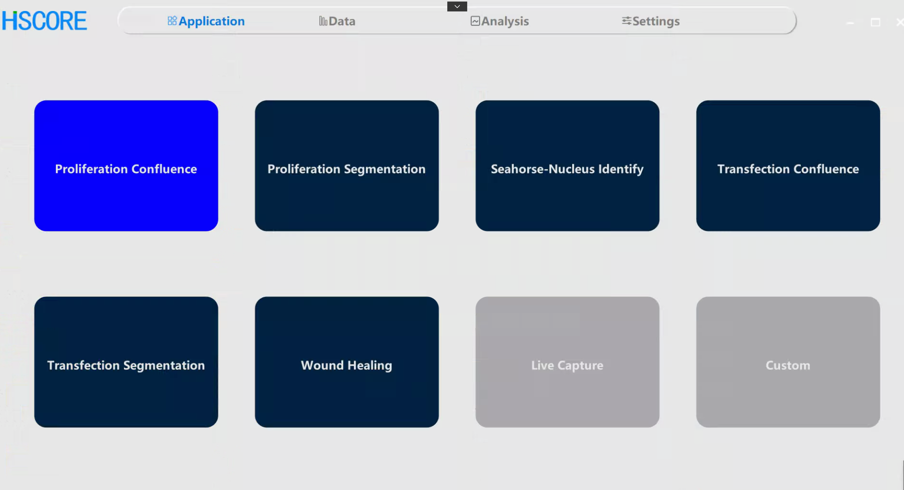
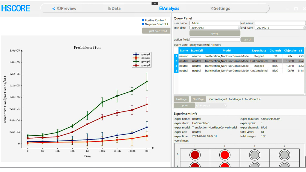
The independent EOS application app is easy to use. The capture view, cycle, and objectives(4x, 10x, 20x, with 40x expansion supported) can all be set on the software without the need for manual adjustment. After the settings are completed, the instrument automatically collects images. The deeply trained AI neural network algorithm of EOS automatically identifies the cell boundaries and generates a curve about the number of cells (/mm2) based on the flow of time .
The EOS Long-term Cell Imaging Workstation is friendly for high-throughput drug screening. It can independently analyze the cell proliferation curves of individual wells in 96- to 384-well plates. The data results are presented in the form of a dataset, enabling compound screening or condition identification in a fast and simple way.
High-precision label-free cell classification and counting
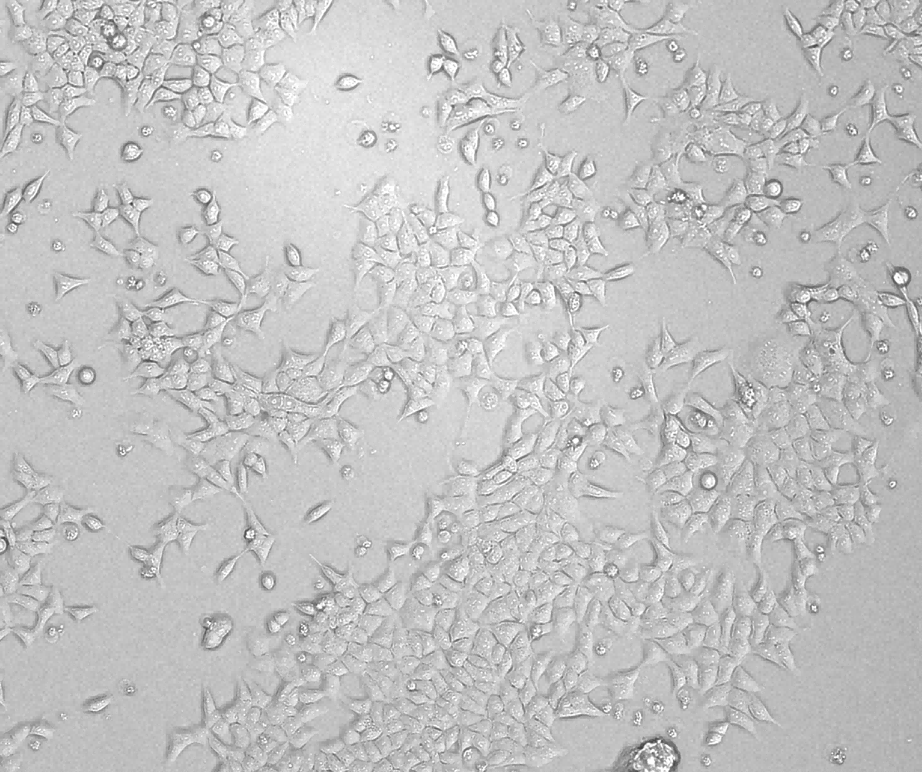
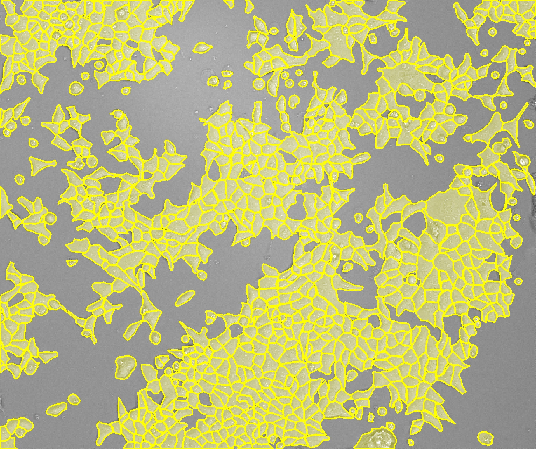
The vast majority of adherent cells have different morphologies. They may present relatively complex shapes such as slender fusiform or synapse-like. At the same time, some semi-adherent and semi-suspended cells are mixed with round suspended cells. Conducting precise label-free classification of these cells is a relatively difficult task. The label-free cell proliferation classification AI algorithm equipped in EOS has undergone in-depth training with a large amount of data from multiple groups of models. It can meet the segmentation requirements of the vast majority of cell types without the need for manual parameter adjustment.
Whole-well capture and analysis
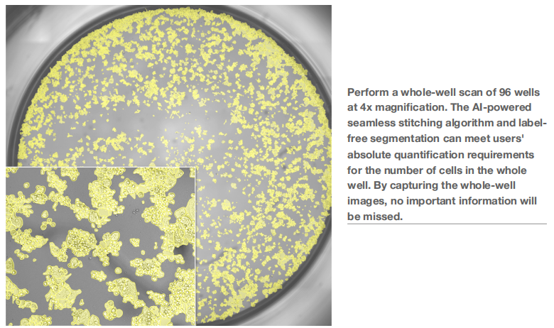
The EOS cell proliferation analysis is not limited to capture at specific locations selected within the well plate. The instrument can be set to the area mode or the whole-well mode to conduct unified analysis of cells in a specific area or the entire well, enabling every cell to realize its value.
Conclusion
The cell proliferation module of the EOS Long-term Cell Imaging Workstation non-invasively and dynamically monitors changes in cell proliferation values based on image algorithms. Through high-precision AI analysis and whole-well/area capture modes, it significantly reduces human operation, improves data accuracy and stability, and eliminates reagent/environmental interference. The neat workflow and high-precision analysis technology offer you a new experience in cell proliferation analysis.